Transcription:
We going to use a cannulated hemi implant for the resurfacing of the 1st MPJ damage by the Arthrodesis or other joint disease.
The patient experience pain in difficulty moving the big toe, the indication are limped into one term Hallux Rigidus. Before we began our demonstration it is useful to become familiar with the surgical tray and the instrumentation of the vilex 1st MPJ system.
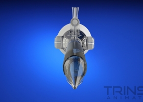
The Kit comes with 2 type of implants concave shape, for the replacement of the base CHI, and Convex shape for the replacement of the head of the meditorcal. The falex end are oval in shape and confirm to the cross section of the falling shaft. They come in 5 sizes, they are available in titanium, cobalt chrome with titanium backing, for faster ossification. The meth head comes in 5 sizes in cobalt chrome with titanium backing only, the vilex MPJ implant offer a number of unique features.
The stem is basically a self drilling and tapping screw requiring no drilling or broaching. The implant is cannulated and screw into the shaft, the driver has 2 prongs that made two holes on the prefer on the implant. The surgical procedure follows make an initial incision dorsal medial dissecting the soft tissue to expose the joint with the free elevator, Elevates the joint to expose the mid head, remove the banian and the dorsal spore as a part of the reshaping meditorcal head, smooth the sharp edges with sign cutting tool, move the curtilage gradually.
starting from the top to expose the trough of the concave surface of base falex. Make a cut between 4 to 5 mm wide to accommodate the implant and decompress the joint, the cut should be neutral perpendicular to the shaft it is permissible to make the cut slidly slided dorsal plantard, to increase the dorsiflexion range of the toe after surgery after exercising the base of the falex, expose the base select the one of the elliptical trial sizes and placed it over exposed surface. The implant should cover as much of the surface as possible with little or no overhang.
Once the correct size is selected run a k-wire to the cannulated seizure and advance the wire about 15mm into the falex and absorb how the seizure is succeeded . It is advisable to use a C-arm to make sure the wire is properly centred. If the wire is not properly positioned remove the wire and reposition it. This is the advantage of the cannulated system, there is no drilling or broaching and thus the positioning of the implant is certain to be correct. Advance the k-wire another 20mm, remove the seizure and slide the corresponding hemi implant over the k-wire and drive the implant with the special 2 prongs driver.
Since the implant is oval in shape it’s initial orientation may be an adequate advance the implant forward as much as one half turn for correct orientation seizure enclosed.
For more information please visit http://www.vilex.com


